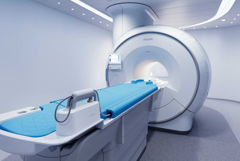
One of the most useful tools in modern medicine is medical imaging. It has changed the way doctors identify, monitor, and treat diseases. It allows doctors to see inside the body without invasive treatments, providing them with vital information that can often save lives. Medical imaging saves lives every day because it helps doctors plan complex treatments and detect deadly diseases early. Medical imaging is now used in almost all hospitals and clinics, making it an essential part of medical practice and improving patient outcomes.
A Game Changer in Discovery and Diagnosis
Before imaging, doctors relied primarily on symptoms, patient reports, and open surgeries to understand what was happening inside the body. This approach was time-consuming, risky, and often led to errors. Everything changed with the advent of diagnostic tools such as X-rays, ultrasounds, CT scans, and MRIs. These tools allow doctors to see inside the body without damaging it and accurately detect health problems. Medical imaging allows doctors to see the disease they are dealing with, often long before symptoms worsen. It can help patients detect tumors, identify broken bones, or check for internal bleeding.
Early Detection of Deadly Diseases
Early detection is one of the most important ways medical imaging saves lives. Today, imaging tools often detect cancer, heart disease, and neurological disorders at an early stage. For example, mammograms help detect breast cancer early, when it is most treatable. Heart and brain scans (MRI) can detect brain tumors even when they are small, as can CT scans. When cancer is detected early, treatment can begin sooner and often with less invasive methods. This helps patients improve their survival and quality of life.
Emergency Care
In emergency care, time is often crucial. In emergency situations such as trauma, stroke, or internal bleeding, medical imaging is crucial to quickly and accurately localizing the condition. When a patient goes to the emergency room with chest pain, they can quickly get an ECG and chest X-ray to make sure there is no heart attack or lung disease. In stroke treatment, a CT or MRI scan can be used immediately to determine if the stroke was caused by a blockage or bleeding in the brain. This helps doctors quickly develop the right treatment plan. These swift actions can determine whether a patient experiences improvement, permanent disability, or even life or death.
Supporting Precision Surgery
Imaging technology is used not only before or after surgery but also during surgery. Real-time imaging helps doctors navigate the body precisely during complex surgeries. MRIs or fluoroscopy instruments used during surgery give surgeons comprehensive visual feedback, allowing them to perform the procedure more safely and efficiently. This increases the chance of successful treatments and reduces the chance of problems. Whether it’s placing a stent in an artery or removing a tumor near a vital organ, imaging helps ensure that treatment is as safe and accurate as possible.
Monitoring Long-Term Conditions
People with chronic conditions need constant monitoring to maintain their health. Imaging tools are essential for this ongoing care. Someone with heart disease may have regular echocardiograms to see how their heart is functioning. Someone with long-term lung disease may have regular chest x-rays or CT scans to monitor changes in their body. Someone with diabetes may need an eye exam to detect signs of diabetic retinopathy. These regular imaging tests help doctors understand when their condition is getting worse and how well their medications are working and quickly adjust their care plans to prevent problems.
Enhancing Maternal and Fetal Health
Imaging is now an important part of prenatal care. Ultrasound allows doctors to closely monitor the growth and development of the fetus during pregnancy. It can detect birth defects, track fetal growth, check the placenta, and ensure that the baby is in the correct position for delivery. This not only gives parents peace of mind but also helps doctors prepare for any problems that may arise before birth. Fetal magnetic resonance imaging (MRI) and other more advanced scanning technologies can be used to better visualize the fetus’ organs during high-risk pregnancies. These tools help make deliveries safer and ensure that mothers and babies are healthier after birth.
Making Cancer Treatment More Accurate
Medical imaging is essential for both detecting cancer and planning treatment. Radiologists use CT scans, MRIs, and PET scans to determine the location, size, and stage of cancer. This information is essential for developing an effective treatment plan. Imaging is used during radiation therapy to ensure that radiation only hits cancer cells and does not damage healthy tissue. We monitor the patient for signs of cancer recurrence after treatment. Being able to visualize cancer at every stage of treatment can make it more effective and significantly increase survival rates.
Conclusion
Medical imaging technology is one of the most important advances in the history of medicine. It gives doctors the tools they need to detect disease early, make accurate diagnoses, perform surgeries more accurately, and monitor treatment outcomes with unprecedented precision. It saves lives every day by reducing guesswork and speeding up and improving medical care. With an X-ray or MRI, doctors can now see inside the body without cutting it open. This breakthrough has revolutionized healthcare. As imaging tools become better, cheaper, and easier to use, their ability to save lives will only increase, making them an essential part of healthcare worldwide.
FAQs
1. Will imaging robots replace doctors?
Absolutely not. Radiologists, physicians, and other trained medical personnel still need to interpret and diagnose the visual information these machines provide.
2. Is imaging used during surgery?
Yes. During surgeries, real-time imaging helps physicians stay focused, improving accuracy and reducing risks in complex surgeries.
3. Is it possible to perform imaging at home?
Most imaging tests are still performed in a clinic or hospital, but some portable ultrasound and mobile imaging devices can be used in home healthcare or other remote areas with the help of a professional.
4. How often should someone get a scan?
It depends on the patient’s health and the doctor’s advice. Some tests, such as bone density scans and mammograms, are performed regularly, depending on age and risk factors.
5. What does the future of medical imaging hold?
In the future, there will be more advanced AI imaging systems, easier access to wearable devices, and better integration with telemedicine, making medical imaging more likely to save lives.